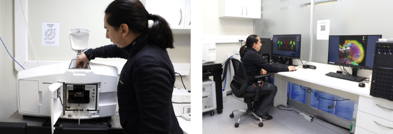
Zeiss Lightsheet 7 Light-Sheet Multiview Imaging of Living and Cleared Specimens
Light sheet fluorescence microscopy (LSFM) is well-suited for rapidly and gently imaging whole living model organisms, tissues, and cells as they develop over extended time periods. The ZEISS Lightsheet 7 microscope enables imaging of large optically cleared specimens in their entirety at subcellular resolution. Specialized optics, sample chambers, and holders allow adapting to the refractive index of the chosen clearing technique.
Lightsheet imaging at http://lightsheetchile.cl
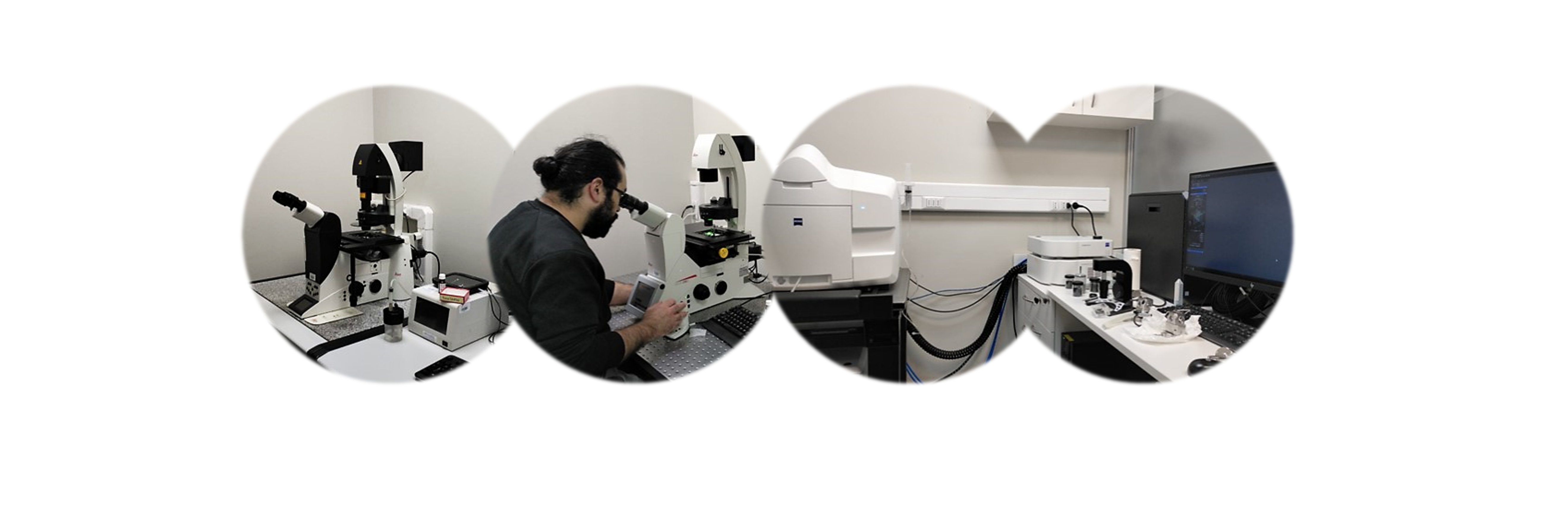
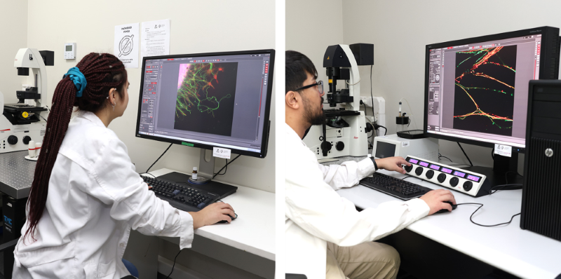
Leica TCS SP8 Confocal Spectral Microscope
The Leica SP8 is an advanced confocal microscopy system designed to provide high-resolution imaging for research applications in life sciences. This microscope excels in its ability to perform fluorescence imaging accurately and efficiently, making it ideal for detailed studies of living cells and tissues.The SP8 features a laser scanning system that allows for image acquisition across multiple wavelengths, facilitating the simultaneous visualization of various fluorescent markers. Its modular design enables the integration of different illumination options and detectors, tailored to the specific needs of each experiment.Additionally, the SP8 is equipped with adaptive focus control, which automatically adjusts focus in real-time, a vital feature for long-term imaging of live cells. Its imaging software, LAS X, offers advanced tools for data collection and analysis, including capabilities for Z-stacking and time-lapse imaging.This combination of flexibility, precision, and advanced imaging capabilities positions the SP8 as an essential tool for cutting-edge scientific research, enabling the easy acquisition of detailed and reproducible data.
Features:
- Spectral confocal detector based on a prism with the capacity for up to 5 confocal PMT detectors (all spectral), each with detection in the range of 400 – 800 nm
- Equipped with 3 confocal PMT detectors.
-VISIR scanner optics compatible with illumination from visible to IR (400 - 1300 nm).
- Maximum digital resolution of 8192 x 8192.
- Scanning speed of 7 fps at 512 x 512 (84 fps at 516 x 16).
- Maximum line scanning frequency of 3600 lines/second.
- Scanning field diameter of 22 mm.
- Hardware confocal zoom from 0.75x to 48x.
- Scanner with 200° optical rotation.
- Transmitted light detector.
- Flexible laser combiner with AOBS technology for maximum transmittance and very specific selection of laser excitation lines, allowing any combination of available laser lines with up to 8 lines simultaneously.
- Available objectives optimized for confocality in the system:
10x/0.30 HC PL FLUOTAR
20x/0.50 HC PL FLUOTAR
40x/0.85 HCX PL APO CORR CS 0.11-0.23
63x/1.40 Oil HC PL APO CS2 immersion objective
- Equipped with Nomarski on all objectives.
- Inverted research microscope Leica DMI6000 B for Confocal SP8 with motorized focus with a minimum step of 50 nm.
- Motorized turret with 6 positions.
- Wide-field fluorescence illumination with a metal halide lamp.
- Fluorescence cubes for initial sample localization: DAPI, FITC, TRICT, and equivalents.
- Manual control mechanical XY stage.
- Motorized tiltable illumination column with S28/0.55 long-distance condenser with halogen illumination.
- LAS X Control Software that allows control of the microscope and image capture in Multi-channel, Time-Lapse, Z-Stack, Lambda-Scan modes, spectral separation, colocalization, creation of complex experiment protocols, acquisition, and analysis of FRET, FLIP, FRAP experiments, and 3D image reconstruction.
- Control computer: High Power HPZ420 Workstation with Windows 7 Professional (64 bit) operating system. - Intel Xeon Quad core E51620 3.6 GHz 10MB 1600- 8 GByte 1600RAM- NVIDIA Quadro K600 1GB high performance GPU- 2 TByte SATA hard disk drive- 16x DVD+/RW Supermulti Drive- eSATA, USB 2.0, IEEE 1394 A/B- Keyboard and mouse- High brilliance 30" LCD (SIPS) flat screen, true color, 2560 x 1600 pixels.
- Passive anti-vibration table.
- Miniature cell incubator for use on the mechanical stage with constant temperature control from ambient +3°-40°C and pre-mixed CO2 at 5%. Includes humidifier, heating ring for immersion objectives, and adapters for: 1) Nunc Lab-Tek II Chamber Slides, 2) Nunc Lab-Tek II Chambered Coverglass, 3) 35mm magnetic culture chamber or 35mm culture plate, 4) Adapter for Well-Slide and Chamlide MB, 5) Chamlide CMB culture chamber for 18mm round coverslips, and 6) Chamlide CMB culture chamber for 25mm round coverslips.
Leica DMi8 Inverted Fluorescence Microscope
The Leica DMi8 microscope is a sophisticated inverted microscope system tailored for research in life sciences, encompassing fields like cell biology and materials science. It features outstanding fluorescence capabilities, supported by high-sensitivity cameras and an effective light management system, which produces bright, clear images while minimizing phototoxicity to living specimens. The DMi8 is equipped with a motorized stage that accommodates various slide formats, from standard slides to multi-well plates, thereby improving throughput for high-content screening tasks.Its imaging software facilitates advanced imaging techniques, including time-lapse, Z-stacking, and multi-channel fluorescence, offering extensive tools for data collection and analysis. Additionally, the DMi8 includes adaptive focus control, which continuously adjusts focus in real-time, a critical feature for long-term live cell imaging. This blend of flexibility, precision, and advanced imaging capabilities positions the DMi8 as an essential instrument for pioneering scientific research, enabling the acquisition of detailed and reproducible data with ease.
Equipped with the following features:
- Motorized focus with a minimum step precision of 50 nm.
- Hardware-mediated autofocus module Adaptive Focus Control (AFC) using an 850 nm IR LED.
- Motorized fluorescence filter turret with 6 positions equipped with 5 filters for: Individual filters for DAPI, GFP, TEXAS RED, and CY5 plus a triple filter DAPI/FITC/TXRED.
- Motorized objective revolver with 6 positions equipped with the following objectives:
5x Mod. HCX PL FLUOTAR 5x/0.15, WD 12.0 mm
10x HC PL FLUOTAR 10x/0.32, WD 11.1 mm
20X Mod. HCX PL FL N 20x/0.40 CORR, WD 3.5 mm
40x Mod. HC PL APO 40X/0.85 CORR, WD 2.21mm
63x Mod. HC PL APO 63x/1.40-0.60 Oil, WD 0.14 mm
- Motorized scanning XY stage with a displacement range of 127 x 83 mm compatible with 160x110mm adapters. Resolution <0.02µm and reproducibility <1µm.
- Universal sample adapter compatible with slides and Petri dishes with diameters of 24 – 68 mm.
- EL6000 metal halide fluorescence system.
- Coded LED transmitted light illumination column.
- Coded S23/0.53 condenser without contrast techniques. Only bright field.
- Motorized 1.6x magnifying lens.
- Cooled monochrome digital camera sCMOS Leica DFC9000 GTC with 4.2 Mgpxl (2048x2048) with a QE of 82% @580 nm - 50 fps - 12/16-bit – pixel size 6.5 µm.
- PC for experiment capture: Leica LAS X Professional Workstation HP Z440 consisting of: Operating System: Win7 Pro 64 Processor: Intel Xeon E5-1620v3 3.5 10M 2133 4C CPU Main Memory: 16GB DDR4-2133 (4x4GB) RegRAM Graphics board: NVIDIA Quadro K620 2GB 1st GFX 1st Hard Drive: 256GB SATA 1st Solid State Drive 2nd Hard drive: 2 TB 7200 RPM SATA 2nd HDD DVD writer: 9.5mm Slim SuperMulti DVDRW 1st ODD Firewire card: IOI PCI Express x1 FW-B 3Port ext.
- LAS X software for control and image capture of the microscope that allows XY capture modes, multi-channel fluorescence capture, Z-stack, Time-lapse, stitching of multiple fields, or multi-location experiments on the sample using the motorized stage, also with the possibility of conducting complex experiments with all available capture modes. Includes 3D visualization module.
- Active anti-vibration table with a perforated surface.